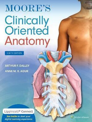
Moore’s Clinically Oriented Anatomy 9th Edition
Moore’s Clinically Oriented Anatomy Ninth Edition( EPUB + Converted PDF ):
North American Edition
Selected as a Doody’s Core Title for 2022!
Lippincott® Connect Featured Title
Purchase of the new print edition of this Lippincott® Connect title includes access to the digital version of the book, plus related materials such as videos and multiple-choice Q&A and self-assessments.
Renowned for its comprehensive coverage and engaging, storytelling approach, the bestselling Moore’s Clinically Oriented Anatomy, 9th Edition, guides students from initial anatomy and foundational science courses through clinical training and practice. A popular resource for a variety of programs, this proven text serves as a complete reference, emphasizing anatomy that is important in physical diagnosis for primary care, interpretation of diagnostic imaging, and understanding the anatomical basis of emergency medicine and general surgery. The 9th Edition reflects the latest changes in the clinical application of anatomy as well as preparation for the USMLE while maintaining the highest standards for scientific and clinical accuracy.
- NEW! Sex and gender content clarifies important gender considerations and reflects an equitable focus on female as well as male anatomy.
- Updated medical imaging and integrated surface anatomy within each chapter clearly demonstrates the relationship between anatomy, physical examination, and diagnosis.
- Extensively revised Clinical Blue Boxes highlight the practical applications of anatomy, accompanied by helpful icons, illustrations, and images that distinguish the type of clinical information covered.
- Updated introductory chapter establishes the foundational understanding of systemic information and basic concepts essential to success from the classroom to the dissection lab.
- Revised comprehensive surface anatomy photographs ensure accurate, effective physical examination diagnoses with integrated natural views of unobstructed surface anatomy and illustrations superimposing anatomical structures with landmarks for more accurate physical examination.
- Insightfully rendered, anatomically accurate illustrations, combined with many photographs and medical images, strengthen comprehension of anatomical concepts and retention of “mental images” of anatomical structures.
- Bottom Line boxes provide detailed summaries at a glance and underscore the “big-picture” perspective.
- Illustrated tables clarify complex information about muscles, veins, arteries, nerves, and other structures for easy study and review.
- Chapter outlines help students find key information quickly and efficiently.
Additional online resources:
- Interactive clinical vignette multiple-choice questions and answers provide valuable self-test opportunities for exam review.
- Case studies engage students in the clinical application process of anatomic principles.
- Clinical Blue Box videos further explore selected clinical applications.
Lippincott® Connect features:
- Full access to the digital version of the book with the ability to highlight and take notes on key passages for a more personal, efficient study experience.
- Carefully curated resources, such as interactive diagrams, audio and video tutorials, and self-assessment, all designed to facilitate further comprehension.
Lippincott® Connect also allows users to create Study Collections to further personalize the study experience. With Study Collections you can:
- Pool content from books across your entire library into self-created Study Collections based on discipline, procedure, organ, concept or other topics.
- Display related text passages, video clips and self-assessment questions from each book (if available) for efficient absorption of material.
- Annotate and highlight key content for easy access later.
- Navigate seamlessly between book chapters, sections, self-assessments, notes and highlights in a single view/page.
Additional ISBNs:
∗ eText ISBN: 1975154096, 978-1975154097, 9781975154097
- See additional information on the Amazon.
More Details
Preface
Acknowledgments
List of Clinical Blue Boxes
List of Tables
Figure Credits
References and Suggested Reading
1 OVERVIEW AND BASIC CONCEPTS
Approaches to Studying Anatomy
Regional Anatomy
Systemic Anatomy
Clinical Anatomy
Sex and Gender
Anatomicomedical Terminology
Anatomical Position
Anatomical Planes
Terms of Relationship and Comparison
Terms of Laterality
Terms of Movement
Anatomical Variations
Integumentary System
Fascias, Fascial Compartments, Bursae, and Potential Spaces
Skeletal System
Cartilage and Bones
Classification of Bones
Bone Markings and Formations
Bone Development
Vasculature and Innervation of Bones
Joints
CLASSIFICATION OF JOINTS
JOINT VASCULATURE AND INNERVATION
Muscle Tissue and Muscular System
Types of Muscle (Muscle Tissue)
Skeletal Muscles
FORM, FEATURES, AND NAMING OF MUSCLES
CONTRACTION OF MUSCLES
FUNCTIONS OF MUSCLES
NERVES AND ARTERIES TO MUSCLES
Cardiac Striated Muscle
Smooth Muscle
Cardiovascular System
Vascular Circuits
Blood Vessels
ARTERIES
VEINS
BLOOD CAPILLARIES
Lymphoid System
Nervous System
Central Nervous System
Peripheral Nervous System
TYPES OF NERVES
SOMATIC AND VISCERAL FIBERS
Somatic Nervous System
Autonomic Nervous System
SYMPATHETIC (THORACOLUMBAR) DIVISION OF AUTONOMIC NERVOUS SYSTEM
PARASYMPATHETIC (CRANIOSACRAL) DIVISION OF AUTONOMIC NERVOUS SYSTEM
ENTERIC NERVOUS SYSTEM
FUNCTIONS OF DIVISIONS OF AUTONOMIC NERVOUS SYSTEM
VISCERAL SENSATION
Medical Imaging Techniques
Conventional Radiography
Computed Tomography
Ultrasonography
Magnetic Resonance Imaging
Nuclear Medicine Imaging
2 BACK
Overview of Back and Vertebral Column
Vertebrae
Structure and Function of Vertebrae
Regional Characteristics of Vertebrae
CERVICAL VERTEBRAE
THORACIC VERTEBRAE
SURFACE ANATOMY OF CERVICAL AND THORACIC VERTEBRAE
LUMBAR VERTEBRAE
SACRUM
COCCYX
SURFACE ANATOMY OF LUMBAR VERTEBRAE, SACRUM, AND COCCYX
Ossification of Vertebrae
Variations in Vertebrae
Vertebral Column
Joints of Vertebral Column
JOINTS OF VERTEBRAL BODIES
JOINTS OF VERTEBRAL ARCHES
ACCESSORY LIGAMENTS OF INTERVERTEBRAL JOINTS
CRANIOVERTEBRAL JOINTS
Movements of Vertebral Column
Curvatures of Vertebral Column
Vasculature of Vertebral Column
Nerves of Vertebral Column
Muscles of Back
Extrinsic Back Muscles
Intrinsic Back Muscles
SUPERFICIAL LAYER
INTERMEDIATE LAYER
DEEP LAYER
PRINCIPAL MUSCLES PRODUCING MOVEMENTS OF INTERVERTEBRAL JOINTS
Surface Anatomy of Back Muscles
Suboccipital and Deep Neck Muscles
Contents of Vertebral Canal
Spinal Cord
Spinal Nerves and Nerve Roots
Spinal Meninges and Cerebrospinal Fluid (CSF)
SPINAL DURA MATER
SPINAL ARACHNOID MATER
SPINAL PIA MATER
SUBARACHNOID SPACE
Vasculature of Spinal Cord and Spinal Nerve Roots
ARTERIES OF SPINAL CORD AND NERVE ROOTS
VEINS OF SPINAL CORD
3 UPPER LIMB
Overview of Upper Limb
Comparison of Upper and Lower Limbs
Bones of Upper Limb
Clavicle
Scapula
Humerus
Bones of Forearm
ULNA
RADIUS
Bones of Hand
OSSIFICATION OF BONES OF HAND
Surface Anatomy of Upper Limb Bones
Fascia of Upper Limb
Vessels and Nerves of Upper Limb
Overview of Arterial Supply of Upper Limb
Venous Drainage of Upper Limb
SUPERFICIAL VEINS OF UPPER LIMB
DEEP VEINS OF UPPER LIMB
Lymphatic Drainage of Upper Limb
Cutaneous and Motor Innervation of Upper Limb
CUTANEOUS INNERVATION OF UPPER LIMB
MOTOR INNERVATION OF UPPER LIMB
Summary of Peripheral Nerves of Upper Limb
Pectoral and Scapular Regions
Anterior Axio-Appendicular Muscles
Posterior Axio-Appendicular and Scapulohumeral Muscles
SUPERFICIAL POSTERIOR AXIO-APPENDICULAR (EXTRINSIC SHOULDER) MUSCLES
DEEP POSTERIOR AXIO-APPENDICULAR (EXTRINSIC SHOULDER) MUSCLES
SCAPULOHUMERAL (INTRINSIC SHOULDER) MUSCLES
ROTATOR CUFF MUSCLES
Surface Anatomy of Pectoral, Scapular, and Deltoid Regions
Axilla
Axillary Artery
Axillary Vein
Axillary Lymph Nodes
Brachial Plexus
Arm
Muscles of Arm
BICEPS BRACHII
BRACHIALIS
CORACOBRACHIALIS
TRICEPS BRACHII
ANCONEUS
Brachial Artery
PROFUNDA BRACHII ARTERY
HUMERAL NUTRIENT ARTERY
SUPERIOR ULNAR COLLATERAL ARTERY
INFERIOR ULNAR COLLATERAL ARTERY
Veins of Arm
SUPERFICIAL VEINS
DEEP VEINS
Nerves of Arm
MUSCULOCUTANEOUS NERVE
RADIAL NERVE
MEDIAN NERVE
ULNAR NERVE
Cubital Fossa
Surface Anatomy of Arm and Cubital Fossa
Forearm
Compartments of Forearm
Muscles of Forearm
FLEXOR–PRONATOR MUSCLES OF FOREARM
EXTENSOR MUSCLES OF FOREARM
Arteries of Forearm
ULNAR ARTERY
RADIAL ARTERY
Veins of Forearm
SUPERFICIAL VEINS
DEEP VEINS
Nerves of Forearm
MEDIAN NERVE IN FOREARM
ULNAR NERVE IN FOREARM
RADIAL NERVE IN FOREARM
LATERAL AND MEDIAL CUTANEOUS NERVES OF FOREARM
Surface Anatomy of Forearm
Hand
Fascia and Compartments of Palm
Muscles of Hand
THENAR MUSCLES
ADDUCTOR POLLICIS
HYPOTHENAR MUSCLES
SHORT MUSCLES OF HAND
Long Flexor Tendons and Tendon Sheaths in Hand
Arteries of Hand
ULNAR ARTERY IN HAND
RADIAL ARTERY IN HAND
Veins of Hand
Nerves of Hand
MEDIAN NERVE IN HAND
ULNAR NERVE IN HAND
RADIAL NERVE IN HAND
Surface Anatomy of Hand
Joints of Upper Limb
Sternoclavicular Joint
ARTICULATION OF STERNOCLAVICULAR JOINT
JOINT CAPSULE OF STERNOCLAVICULAR JOINT
LIGAMENTS OF STERNOCLAVICULAR JOINT
MOVEMENTS OF STERNOCLAVICULAR JOINT
BLOOD SUPPLY OF STERNOCLAVICULAR JOINT
NERVE SUPPLY OF STERNOCLAVICULAR JOINT
Acromioclavicular Joint
ARTICULATION OF ACROMIOCLAVICULAR JOINT
JOINT CAPSULE OF ACROMIOCLAVICULAR JOINT
LIGAMENTS AUGMENTING ACROMIOCLAVICULAR JOINT
MOVEMENTS OF ACROMIOCLAVICULAR JOINT
BLOOD SUPPLY OF ACROMIOCLAVICULAR JOINT
NERVE SUPPLY OF ACROMIOCLAVICULAR JOINT
Glenohumeral Joint
ARTICULATION OF GLENOHUMERAL JOINT
JOINT CAPSULE OF GLENOHUMERAL JOINT
LIGAMENTS OF GLENOHUMERAL JOINT
MOVEMENTS OF GLENOHUMERAL JOINT
MUSCLES MOVING GLENOHUMERAL JOINT
BLOOD SUPPLY OF GLENOHUMERAL JOINT
INNERVATION OF GLENOHUMERAL JOINT
BURSAE AROUND GLENOHUMERAL JOINT
SUBTENDINOUS BURSA OF SUBSCAPULARIS
SUBACROMIAL BURSA
Elbow Joint
ARTICULATION OF ELBOW JOINT
JOINT CAPSULE OF ELBOW JOINT
LIGAMENTS OF ELBOW JOINT
MOVEMENTS OF ELBOW JOINT
MUSCLES MOVING ELBOW JOINT
BLOOD SUPPLY OF ELBOW JOINT
NERVE SUPPLY OF ELBOW JOINT
BURSAE AROUND ELBOW JOINT
Proximal Radio-Ulnar Joint
ARTICULATION OF PROXIMAL RADIO-ULNAR JOINT
JOINT CAPSULE OF PROXIMAL RADIO-ULNAR JOINT
LIGAMENTS OF PROXIMAL RADIO-ULNAR JOINT
MOVEMENTS OF PROXIMAL RADIO-ULNAR JOINT
MUSCLES MOVING PROXIMAL RADIO-ULNAR JOINT
BLOOD SUPPLY OF PROXIMAL RADIO-ULNAR JOINT
INNERVATION OF PROXIMAL RADIO-ULNAR JOINT
Distal Radio-Ulnar Joint
ARTICULATION OF DISTAL RADIO-ULNAR JOINT
JOINT CAPSULE OF DISTAL RADIO-ULNAR JOINT
LIGAMENTS OF DISTAL RADIO-ULNAR JOINT
MOVEMENTS OF DISTAL RADIO-ULNAR JOINT
MUSCLES MOVING DISTAL RADIO-ULNAR JOINT
BLOOD SUPPLY OF DISTAL RADIO-ULNAR JOINT
INNERVATION OF DISTAL RADIO-ULNAR JOINT
Wrist Joint
ARTICULATION OF WRIST JOINT
JOINT CAPSULE OF WRIST JOINT
LIGAMENTS OF WRIST JOINT
MOVEMENTS OF WRIST JOINT
MUSCLES MOVING WRIST JOINT
BLOOD SUPPLY OF WRIST JOINT
INNERVATION OF WRIST JOINT
Intercarpal Joints
JOINT CAPSULE OF INTERCARPAL JOINTS
LIGAMENTS OF INTERCARPAL JOINTS
MOVEMENTS OF INTERCARPAL JOINTS
BLOOD SUPPLY OF INTERCARPAL JOINTS
INNERVATION OF INTERCARPAL JOINTS
Carpometacarpal and Intermetacarpal Joints
ARTICULATIONS OF CARPOMETACARPAL AND INTERMETACARPAL JOINTS
JOINT CAPSULE OF CARPOMETACARPAL AND INTERMETACARPAL JOINTS
LIGAMENTS OF CARPOMETACARPAL AND INTERMETACARPAL JOINTS
MOVEMENTS OF CARPOMETACARPAL AND INTERMETACARPAL JOINTS
BLOOD SUPPLY OF CARPOMETACARPAL AND INTERMETACARPAL JOINTS
INNERVATION OF CARPOMETACARPAL AND INTERMETACARPAL JOINTS
Metacarpophalangeal and Interphalangeal Joints
ARTICULATIONS OF METACARPOPHALANGEAL AND INTERPHALANGEAL JOINTS
JOINT CAPSULES OF METACARPOPHALANGEAL AND INTERPHALANGEAL JOINTS
LIGAMENTS OF METACARPOPHALANGEAL AND INTERPHALANGEAL JOINTS
MOVEMENTS OF METACARPOPHALANGEAL AND INTERPHALANGEAL JOINTS
BLOOD SUPPLY OF METACARPAL AND INTERPHALANGEAL JOINTS
INNERVATION OF METACARPAL AND INTERPHALANGEAL JOINTS
4 THORAX
Overview of Thorax
Thoracic Wall
Skeleton of Thoracic Wall
RIBS, COSTAL CARTILAGES, AND INTERCOSTAL SPACES
THORACIC VERTEBRAE
STERNUM
Thoracic Apertures
SUPERIOR THORACIC APERTURE
INFERIOR THORACIC APERTURE
Joints of Thoracic Wall
COSTOVERTEBRAL JOINTS
STERNOCOSTAL JOINTS
Movements of Thoracic Wall
Muscles of Thoracic Wall
Fascia of Thoracic Wall
Nerves of Thoracic Wall
TYPICAL INTERCOSTAL NERVES
ATYPICAL INTERCOSTAL NERVES
Vasculature of Thoracic Wall
ARTERIES OF THORACIC WALL
VEINS OF THORACIC WALL
Breasts
FEMALE BREASTS
VASCULATURE OF BREAST
NERVES OF BREAST
Surface Anatomy of Thoracic Wall
Viscera of Thoracic Cavity
Pleurae, Lungs, and Tracheobronchial Tree
PLEURAE
LUNGS
TRACHEOBRONCHIAL TREE
VASCULATURE OF LUNGS AND PLEURAE
NERVES OF LUNGS AND PLEURAE
SURFACE ANATOMY OF PLEURAE AND LUNGS
Overview of Mediastinum
Pericardium
Heart
RIGHT ATRIUM
RIGHT VENTRICLE
LEFT ATRIUM
LEFT VENTRICLE
SEMILUNAR VALVES
VASCULATURE OF HEART
STIMULATING, CONDUCTING, AND REGULATING SYSTEMS OF HEART
Superior Mediastinum and Great Vessels
THYMUS
GREAT VESSELS
NERVES IN SUPERIOR MEDIASTINUM
TRACHEA
ESOPHAGUS
Posterior Mediastinum
THORACIC AORTA
ESOPHAGUS
THORACIC DUCT AND LYMPHATIC TRUNKS
VESSELS AND LYMPH NODES OF POSTERIOR MEDIASTINUM
NERVES OF POSTERIOR MEDIASTINUM
Anterior Mediastinum
5 ABDOMEN
Overview: Walls, Cavities, Regions, and Planes
Anterolateral Abdominal Wall
Fascia of Anterolateral Abdominal Wall
Muscles of Anterolateral Abdominal Wall
EXTERNAL OBLIQUE MUSCLE
INTERNAL OBLIQUE MUSCLE
TRANSVERSUS ABDOMINIS MUSCLE
RECTUS ABDOMINIS MUSCLE
PYRAMIDALIS
RECTUS SHEATH, LINEA ALBA, AND UMBILICAL RING
FUNCTIONS AND ACTIONS OF ANTEROLATERAL ABDOMINAL MUSCLES
Neurovasculature of Anterolateral Abdominal Wall
DERMATOMES OF ANTEROLATERAL ABDOMINAL WALL
NERVES OF ANTEROLATERAL ABDOMINAL WALL
VESSELS OF ANTEROLATERAL ABDOMINAL WALL
Internal Surface of Anterolateral Abdominal Wall
Inguinal Region
INGUINAL LIGAMENT AND ILIOPUBIC TRACT
INGUINAL CANAL
Spermatic Cord, Scrotum, and Testes
SPERMATIC CORD
SCROTUM
TESTES
EPIDIDYMIS
Surface Anatomy of Anterolateral Abdominal Wall
Peritoneum and Peritoneal Cavity
Embryology of Peritoneal Cavity
Peritoneal Formations
Subdivisions of Peritoneal Cavity
Abdominal Viscera
Overview of Abdominal Viscera and Digestive Tract
Esophagus
Stomach
POSITION, PARTS, AND SURFACE PROJECTION OF STOMACH
INTERIOR OF STOMACH
RELATIONS OF STOMACH
VESSELS AND NERVES OF STOMACH
Small Intestine
DUODENUM
JEJUNUM AND ILEUM
Large Intestine
CECUM AND APPENDIX
COLON
RECTUM AND ANAL CANAL
Spleen
Pancreas
Liver
SURFACE PROJECTION, SURFACES, PERITONEAL REFLECTIONS, AND RELATIONSHIPS OF LIVER
ANATOMICAL LOBES OF LIVER
FUNCTIONAL SUBDIVISION OF LIVER
BLOOD VESSELS OF LIVER
LYMPHATIC DRAINAGE AND INNERVATION OF LIVER
Biliary Ducts and Gallbladder
BILE DUCT
GALLBLADDER
HEPATIC PORTAL VEIN AND PORTAL–SYSTEMIC ANASTOMOSES
Kidneys, Ureters, and Suprarenal Glands
KIDNEYS
URETERS
SUPRARENAL GLANDS
VESSELS AND NERVES OF KIDNEYS, URETERS, AND SUPRARENAL GLANDS
Innervation of Abdominal Viscera
SYMPATHETIC INNERVATION
PARASYMPATHETIC INNERVATION
EXTRINSIC AUTONOMIC PLEXUSES
INTRINSIC PLEXUSES: THE ENTERIC NERVOUS SYSTEM
VISCERAL SENSORY INNERVATION
Diaphragm
Vessels and Nerves of Diaphragm
Diaphragmatic Apertures
CAVAL OPENING
ESOPHAGEAL HIATUS
AORTIC HIATUS
SMALL OPENINGS IN DIAPHRAGM
Actions of Diaphragm
Posterior Abdominal Wall
Fascia of Posterior Abdominal Wall
Muscles of Posterior Abdominal Wall
PSOAS MAJOR
ILIACUS
QUADRATUS LUMBORUM
Nerves of Posterior Abdominal Wall
Vessels of Posterior Abdominal Wall
ABDOMINAL AORTA
VEINS OF POSTERIOR ABDOMINAL WALL
LYMPHATIC VESSELS AND LYMPH NODES OF POSTERIOR ABDOMINAL WALL
Sectional Medical Imaging of Abdomen
6 PELVIS AND PERINEUM
Introduction to Pelvis and Perineum
Pelvic Girdle
Bones and Features of Pelvic Girdle
Orientation of Pelvic Girdle
Pelvic Girdle Sexual Differences
Joints and Ligaments of Pelvic Girdle
SACRO-ILIAC JOINTS
PUBIC SYMPHYSIS
LUMBOSACRAL JOINTS
SACROCOCCYGEAL JOINT
Pelvic Cavity
Walls and Floor of Pelvic Cavity
ANTERO-INFERIOR PELVIC WALL
LATERAL PELVIC WALLS
POSTERIOR WALL (POSTEROLATERAL WALL AND ROOF)
PELVIC FLOOR
Peritoneum and Peritoneal Cavity of Pelvis
Pelvic Fascia
MEMBRANOUS PELVIC FASCIA: PARIETAL AND VISCERAL
ENDOPELVIC FASCIA: LOOSE AND CONDENSED
Neurovascular Structures of Pelvis
Pelvic Arteries
INTERNAL ILIAC ARTERY
OVARIAN ARTERY
MEDIAN SACRAL ARTERY
SUPERIOR RECTAL ARTERY
Pelvic Veins
Lymph Nodes of Pelvis
Pelvic Nerves
OBTURATOR NERVE
LUMBOSACRAL TRUNK
SACRAL PLEXUS
COCCYGEAL PLEXUS
PELVIC AUTONOMIC NERVES
VISCERAL AFFERENT INNERVATION IN PELVIS
Pelvic Viscera
Urinary Organs
URETERS
PROXIMAL (PELVIC) MALE URETHRA
FEMALE URETHRA
Rectum
ARTERIAL SUPPLY AND VENOUS DRAINAGE OF RECTUM
INNERVATION OF RECTUM
Male Internal Genital Organs
DUCTUS DEFERENS
SEMINAL GLANDS
EJACULATORY DUCTS
PROSTATE
BULBO-URETHRAL GLANDS
INNERVATION OF INTERNAL GENITAL ORGANS OF MALE PELVIS
Female Internal Genital Organs
OVARIES
UTERINE TUBES
UTERUS
VAGINA
ARTERIAL SUPPLY AND VENOUS DRAINAGE OF VAGINA
INNERVATION OF VAGINA AND UTERUS
Lymphatic Drainage of Pelvic Viscera
LYMPHATIC DRAINAGE FROM URINARY SYSTEM
LYMPHATIC DRAINAGE FROM RECTUM
LYMPHATIC DRAINAGE FROM MALE PELVIC VISCERA
LYMPHATIC DRAINAGE FROM FEMALE PELVIC VISCERA
Perineum
Fasciae and Pouches of Urogenital Triangle
PERINEAL FASCIAE
SUPERFICIAL PERINEAL POUCH
DEEP PERINEAL POUCH
Features of Anal Triangle
ISCHIO-ANAL FOSSAE
PUDENDAL CANAL AND ITS NEUROVASCULAR BUNDLE
ANAL CANAL
Male Urogenital Triangle
DISTAL MALE URETHRA
SCROTUM
PENIS
LYMPHATIC DRAINAGE OF MALE PERINEUM
PERINEAL MUSCLES OF MALE
ERECTION, EMISSION, EJACULATION, AND REMISSION
Female Urogenital Triangle
FEMALE EXTERNAL GENITALIA
LYMPHATIC DRAINAGE OF FEMALE PERINEUM
PERINEAL MUSCLES OF FEMALE
Sectional Imaging of Pelvis and Perineum
Magnetic Resonance Imaging
7 LOWER LIMB
Overview of Lower Limb
Development of Lower Limb
Bones of Lower Limb
Arrangement of Lower Limb Bones
Hip Bone
ILIUM
ISCHIUM
PUBIS
OBTURATOR FORAMEN
ACETABULUM
ANATOMICAL POSITION OF HIP BONE
Femur
SURFACE ANATOMY OF PELVIC GIRDLE AND FEMUR
Patella
Tibia and Fibula
TIBIA
FIBULA
SURFACE ANATOMY OF TIBIA AND FIBULA
Bones of Foot
TARSUS
METATARSUS
PHALANGES
SURFACE ANATOMY OF BONES OF FOOT
Fascia of Lower Limb
Subcutaneous Tissue
Deep Fascia
FASCIA LATA
DEEP FASCIA OF LEG
Overview of VEssels and Nerves of Lower Limb
Arterial Supply of Lower Limb
Venous Drainage of Lower Limb
SUPERFICIAL VEINS OF LOWER LIMB
DEEP VEINS OF LOWER LIMB
Lymphatic Drainage of Lower Limb
Cutaneous Innervation of Lower Limb
Motor Innervation of Lower Limb
Peripheral Nerves of Lower Limb
Posture and Gait
Standing at Ease
Walking: Gait Cycle
Anterior and Medial Regions of Thigh
Organization of Proximal Lower Limb
Anterior Thigh Muscles
PECTINEUS
ILIOPSOAS
SARTORIUS
QUADRICEPS FEMORIS
Medial Thigh Muscles
ADDUCTOR LONGUS
ADDUCTOR BREVIS
ADDUCTOR MAGNUS
GRACILIS
OBTURATOR EXTERNUS
ACTIONS OF ADDUCTOR MUSCLE GROUP
ADDUCTOR HIATUS
Neurovascular Structures and Relationships in Anteromedial Thigh
FEMORAL TRIANGLE
FEMORAL NERVE
FEMORAL SHEATH
FEMORAL ARTERY
FEMORAL VEIN
ADDUCTOR CANAL
Surface Anatomy of Anterior and Medial Regions of Thigh
Gluteal and Posterior Thigh Regions
Gluteal Region: Buttocks and Hip Region
GLUTEAL LIGAMENTS
Muscles of Gluteal Region
GLUTEUS MAXIMUS
GLUTEUS MEDIUS AND GLUTEUS MINIMUS
TENSOR FASCIAE LATAE
PIRIFORMIS
OBTURATOR INTERNUS AND GEMELLI
QUADRATUS FEMORIS
OBTURATOR EXTERNUS
Posterior Thigh Region
SEMITENDINOSUS
SEMIMEMBRANOSUS
BICEPS FEMORIS
Neurovascular Structures of Gluteal and Posterior Thigh Regions
CLUNIAL NERVES
DEEP GLUTEAL NERVES
ARTERIES OF GLUTEAL AND POSTERIOR THIGH REGIONS
VEINS OF GLUTEAL AND POSTERIOR THIGH REGIONS
LYMPHATIC DRAINAGE OF GLUTEAL AND THIGH REGIONS
Surface Anatomy of Gluteal and Posterior Thigh Regions
Popliteal Fossa and Leg
Popliteal Region
FASCIA OF POPLITEAL FOSSA
NEUROVASCULAR STRUCTURES AND RELATIONSHIPS IN POPLITEAL FOSSA
Anterior Compartment of Leg
ORGANIZATION OF LEG
MUSCLES OF ANTERIOR COMPARTMENT OF LEG
NERVE OF ANTERIOR COMPARTMENT OF LEG
ARTERY IN ANTERIOR COMPARTMENT OF LEG
Lateral Compartment of Leg
MUSCLES IN LATERAL COMPARTMENT OF LEG
NERVES IN LATERAL COMPARTMENT OF LEG
BLOOD VESSELS IN LATERAL COMPARTMENT OF LEG
Posterior Compartment of Leg
SUPERFICIAL MUSCLE GROUP IN POSTERIOR COMPARTMENT
DEEP MUSCLE GROUP IN POSTERIOR COMPARTMENT
NERVES IN POSTERIOR COMPARTMENT
ARTERIES IN POSTERIOR COMPARTMENT
Surface Anatomy of Leg
Foot
Skin and Fascia of Foot
SKIN AND SUBCUTANEOUS TISSUE
DEEP FASCIA OF FOOT
Muscles of Foot
Neurovascular Structures and Relationships in Foot
NERVES OF FOOT
ARTERIES OF FOOT
ARTERIES OF SOLE OF FOOT
VENOUS DRAINAGE OF FOOT
LYMPHATIC DRAINAGE OF FOOT
Surface Anatomy of Ankle and Foot Regions
Joints of Lower Limb
Hip Joint
ARTICULAR SURFACES OF HIP JOINT
JOINT CAPSULE OF HIP JOINT
MOVEMENTS OF HIP JOINT
BLOOD SUPPLY OF HIP JOINT
NERVE SUPPLY OF HIP JOINT
Knee Joint
ARTICULATIONS, ARTICULAR SURFACES, AND STABILITY OF KNEE JOINT
JOINT CAPSULE OF KNEE JOINT
EXTRACAPSULAR LIGAMENTS OF KNEE JOINT
INTRA-ARTICULAR LIGAMENTS OF KNEE JOINT
MOVEMENTS OF KNEE JOINT
BLOOD SUPPLY OF KNEE JOINT
INNERVATION OF KNEE JOINT
BURSAE AROUND KNEE JOINT
Tibiofibular Joints
TIBIOFIBULAR JOINT
TIBIOFIBULAR SYNDESMOSIS
Ankle Joint
ARTICULAR SURFACES OF ANKLE JOINT
JOINT CAPSULE OF ANKLE JOINT
LIGAMENTS OF ANKLE JOINT
MOVEMENTS OF ANKLE JOINT
BLOOD SUPPLY OF ANKLE JOINT
NERVE SUPPLY OF ANKLE JOINT
Foot Joints
MAJOR LIGAMENTS OF FOOT
ARCHES OF FOOT
Surface Anatomy of Joints of Knee, Ankle, and Foot
8 HEAD
Overview of Head
Cranium
Anterior Aspect of Cranium
Lateral Aspect of Cranium
Occipital Aspect of Cranium
Superior Aspect of Cranium
External Surface of Cranial Base
Internal Surface of Cranial Base
ANTERIOR CRANIAL FOSSA
MIDDLE CRANIAL FOSSA
POSTERIOR CRANIAL FOSSA
Walls of Cranial Cavity
Regions of Head
Face and Scalp
Face
Scalp
Muscles of Face and Scalp
MUSCLES OF SCALP, FOREHEAD, AND EYEBROWS
MUSCLES OF MOUTH, LIPS, AND CHEEKS
MUSCLES OF ORBITAL OPENING
MUSCLES OF NOSE AND EARS
Nerves of Face and Scalp
CUTANEOUS NERVES OF FACE AND SCALP
OPHTHALMIC NERVE
MAXILLARY NERVE
MANDIBULAR NERVE
NERVES OF SCALP
MOTOR NERVES OF FACE
FACIAL NERVE
Superficial Vasculature of Face and Scalp
SUPERFICIAL ARTERIES OF FACE
ARTERIES OF SCALP
EXTERNAL VEINS OF FACE
VEINS OF SCALP
LYMPHATIC DRAINAGE OF FACE AND SCALP
Surface Anatomy of Face
Cranial Meninges
Dura Mater
DURAL INFOLDINGS OR REFLECTIONS
DURAL VENOUS SINUSES
VASCULATURE OF DURA MATER
NERVE SUPPLY OF DURA MATER
Arachnoid Mater and Pia Mater
Meningeal Spaces
Brain
Parts of Brain
Ventricular System of Brain
VENTRICLES OF BRAIN
SUBARACHNOID CISTERNS
SECRETION OF CEREBROSPINAL FLUID
CIRCULATION OF CEREBROSPINAL FLUID
ABSORPTION OF CEREBROSPINAL FLUID
FUNCTIONS OF CEREBROSPINAL FLUID
Arterial Blood Supply to Brain
INTERNAL CAROTID ARTERIES
VERTEBRAL ARTERIES
CEREBRAL ARTERIES
CEREBRAL ARTERIAL CIRCLE
Venous Drainage of Brain
Orbits, Eyeball, and Accessory Visual Structures
Orbits
Anterior Accessory Visual Structures
EYELIDS
LACRIMAL APPARATUS
Eyeball
FIBROUS LAYER OF EYEBALL
VASCULAR LAYER OF EYEBALL
INNER LAYER OF EYEBALL
REFRACTIVE MEDIA AND COMPARTMENTS OF EYEBALL
Extra-Ocular Muscles of Orbit
LEVATOR PALPEBRAE SUPERIORIS
MOVEMENTS OF EYEBALL
RECTI AND OBLIQUE MUSCLES
SUPPORTING APPARATUS OF EYEBALL
Nerves of Orbit
Vasculature of Orbit
ARTERIES OF ORBIT
VEINS OF ORBIT
Surface Anatomy of Eye and Lacrimal Apparatus
Parotid and Temporal Regions, Infratemporal Fossa, and Temporomandibular Joint
Parotid Region
PAROTID GLAND
INNERVATION OF PAROTID GLAND AND RELATED STRUCTURES
Temporal Region
Infratemporal Fossa
TEMPOROMANDIBULAR JOINT
MUSCLES OF MASTICATION
NEUROVASCULATURE OF INFRATEMPORAL FOSSA
Oral Region
Oral Cavity
Lips, Cheeks, and Gingivae
LIPS AND CHEEKS
GINGIVAE
Teeth
PARTS AND STRUCTURE OF TEETH
VASCULATURE OF TEETH
INNERVATION OF TEETH
Palate
HARD PALATE
SOFT PALATE
SUPERFICIAL FEATURES OF PALATE
MUSCLES OF SOFT PALATE
VASCULATURE AND INNERVATION OF PALATE
Tongue
PARTS AND SURFACES OF TONGUE
MUSCLES OF TONGUE
INNERVATION OF TONGUE
VASCULATURE OF TONGUE
Salivary Glands
SUBMANDIBULAR GLANDS
SUBLINGUAL GLANDS
Pterygopalatine Fossa
Pterygopalatine Part of Maxillary Artery
Maxillary Nerve
Nose
External Nose
SKELETON OF EXTERNAL NOSE
NASAL SEPTUM
Nasal Cavities
BOUNDARIES OF NASAL CAVITIES
FEATURES OF NASAL CAVITIES
Vasculature and Innervation of Nose
Paranasal Sinuses
FRONTAL SINUSES
ETHMOIDAL CELLS
SPHENOIDAL SINUSES
MAXILLARY SINUSES
Ear
External Ear
AURICLE
EXTERNAL ACOUSTIC MEATUS AND TYMPANIC MEMBRANE
Middle Ear
WALLS OF TYMPANIC CAVITY
PHARYNGOTYMPANIC TUBE
AUDITORY OSSICLES
Internal Ear
BONY LABYRINTH
MEMBRANOUS LABYRINTH
INTERNAL ACOUSTIC MEATUS
9 NECK
Overview
Bones of Neck
Cervical Vertebrae
Hyoid Bone
Fascia of Neck
Cervical Subcutaneous Tissue and Platysma
PLATYSMA
Deep Cervical Fascia
INVESTING LAYER OF DEEP CERVICAL FASCIA
PRETRACHEAL LAYER OF DEEP CERVICAL FASCIA
PREVERTEBRAL LAYER OF DEEP CERVICAL FASCIA
Superficial Structures of Neck: Cervical Regions
Sternocleidomastoid Region
Posterior Cervical Region
Lateral Cervical Region
MUSCLES IN LATERAL CERVICAL REGION
ARTERIES IN LATERAL CERVICAL REGION
VEINS IN LATERAL CERVICAL REGION
NERVES OF LATERAL CERVICAL REGION
LYMPH NODES IN LATERAL CERVICAL REGION
Anterior Cervical Region
MUSCLES IN ANTERIOR CERVICAL REGION
ARTERIES IN ANTERIOR CERVICAL REGION
VEINS IN ANTERIOR CERVICAL REGION
NERVES IN ANTERIOR CERVICAL REGION
Surface Anatomy of Cervical Regions and Triangles of Neck
Deep Structures of Neck
Prevertebral Muscles
Root of Neck
ARTERIES IN ROOT OF NECK
VEINS IN ROOT OF NECK
NERVES IN ROOT OF NECK
Viscera of Neck
Endocrine Layer of Cervical Viscera
THYROID GLAND
PARATHYROID GLANDS
Respiratory Layer of Cervical Viscera
LARYNX
TRACHEA
Alimentary Layer of Cervical Viscera
PHARYNX
ESOPHAGUS
Surface Anatomy of Endocrine and Respiratory Layers of Cervical Viscera
Lymphatics of Neck
10 SUMMARY OF CRANIAL NERVES
Overview
Olfactory Nerve (CN I)
Optic Nerve (CN II)
Oculomotor Nerve (CN III)
Trochlear Nerve (CN IV)
Trigeminal Nerve (CN V)
Ophthalmic Nerve (CN V1)
Maxillary Nerve (CN V2)
Mandibular Nerve (CN V3)
Abducent Nerve (CN VI)
Facial Nerve (CN VII)
Somatic (Branchial) Motor
Visceral (Parasympathetic) Motor
Somatic (General) Sensory
Special Sensory (Taste)
Vestibulocochlear Nerve (CN VIII)
Glossopharyngeal Nerve (CN IX)
Somatic (Branchial) Motor
Visceral (Parasympathetic) Motor
Somatic (General) Sensory
Special Sensory (Taste)
Visceral Sensory
Vagus Nerve (CN X)
Spinal Accessory Nerve (CN XI)
Hypoglossal Nerve (CN XII)
INDEX





















 Dentistry
Reviews
There are no reviews yet.