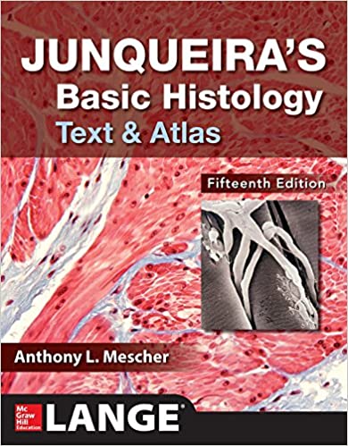
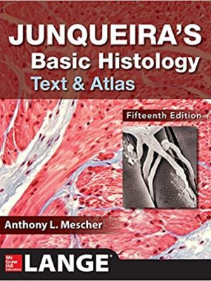
Junqueira’s Basic Histology: Text and Atlas Fifteenth Edition (15th Edition)
The text that has defined histology for generations―concise, clear, beautifully illustrated, and better than ever
A Doody’s Core Title for 2019!
For more than four decades, Junqueira’s Basic Histology has built a global reputation as the most accessible, yet comprehensive overview of human tissue structure and function available. This trusted classic delivers a well-organized and concise presentation of cell biology and histology that integrates the material with that of biochemistry, immunology, endocrinology, and physiology, and provides an excellent foundation for subsequent studies in pathology. Junqueira’s is written specifically for students of medicine and other health-related professions, as well as for advanced undergraduate courses in tissue biology – and there is nothing else like it.
FEATURES
• Electron and light micrographs comprise a definitive atlas of cell, tissue, and organ structures
• NEW! Each chapter now includes a set of multiple-choice self-test questions that allow you to assess your comprehension of important material, with some questions utilizing clinical vignettes or cases to provide real-world relevance
• Summary of Key Points and Summary Tables highlight what is important and present it in a way that makes it memorable
• Streamlined page design ―including concise, high-yield paragraphs; bullets; and bolded key terms
• Acclaimed art and other figures facilitate learning and visualization of key aspects of cell biology and histology
• A cohesive organization examines how to study the structures of cells and tissues; the cell cytoplasm and nucleus; and the four basic tissue types and their role in the organ systems
• Clinical Correlations presented with each topic
• All-inclusive coverage encompasses all tissues, every organ system, organs, bone and cartilage, blood, skin, and more
• Valuable appendix on light microscopy stains clearly explains this need-to-know staining technique
About the Author
Anthony L. Mescher, PhD is Professor of Anatomy and Cell Biology at the Indiana University School of Medicine.
Table of Contents
Chapter 1: Histology & Its Methods of Study
Chapter 2: The Cytoplasm
Chapter 3: The Nucleus
Chapter 4: Epithelial Tissue
Chapter 5: Connective Tissue
Chapter 6: Adipose Tissue
Chapter 7: Cartilage
Chapter 8: Bone
Chapter 9: Nerve Tissue & the Nervous System
Chapter 10: Muscle Tissue
Chapter 11: The Circulatory System
Chapter 12: Blood
Chapter 13: Hemopoiesis
Chapter 14: The Immune System & Lymphoid Organs
Chapter 15: Digestive Tract
Chapter 16: Organs Associated with the Digestive Tract
Chapter 17: The Respiratory System
Chapter 18: Skin
Chapter 19: The Urinary System
Chapter 20: Endocrine Glands
Chapter 21: The Male Reproductive System
Chapter 22: The Female Reproductive System
Chapter 23: The Eye & Ear: Special Sense Organs
Appendix: Light Microscopy Stains








 Dentistry
Reviews
There are no reviews yet.